Notice what helps you.
Try cutting back on coffee and alcohol, and avoid drinking lots of fluids right before bed. These changes can ease the discomfort.
Visual journey · Prostate
Why can it be hard to urinate even without cancer?
The prostate sits in the pelvis, the lowest part of the torso.
Scroll to get closer
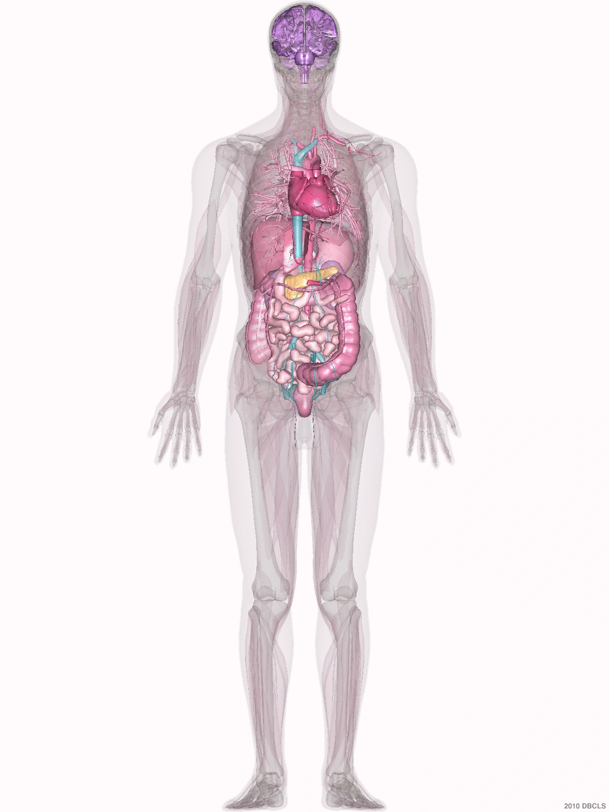
Inside the pelvis
It makes some of the fluid in semen. Running through it is the urethra, the tube that carries urine out of the body.
See what happens when it grows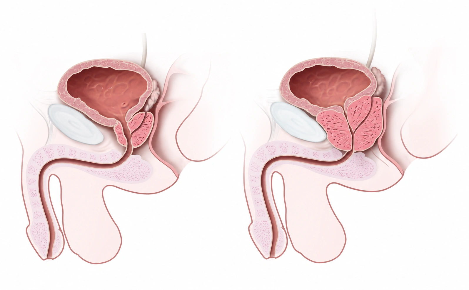 BladderProstateUrethra
Benign enlargement
With age, the prostate can grow larger without cancer. This is called benign prostatic hyperplasia. When it squeezes the urethra, it can weaken your stream or make it hard to start urinating.
BladderProstateUrethraUrine leaves through hereBladderEnlarged prostateNarrower passagein the urethraFollow the urethra down from the bladder: it passes through the prostate before continuing out of the body.
Now look at the same spot: the enlarged tissue squeezes the tube that urine has to pass through.
Other causes, such as an infection, can also make urinating difficult. Your doctor can find out which one it is.
Benign enlargement is not a stage of cancer.
To understand the difference, let’s look at how prostate tissue is organized.
Inside the prostate
This acinus, one of the prostate’s tiny glands, has been cut open to show the inside. Its cells surround the space where its secretions collect.
That space is not the urethra, the tube urine passes through.
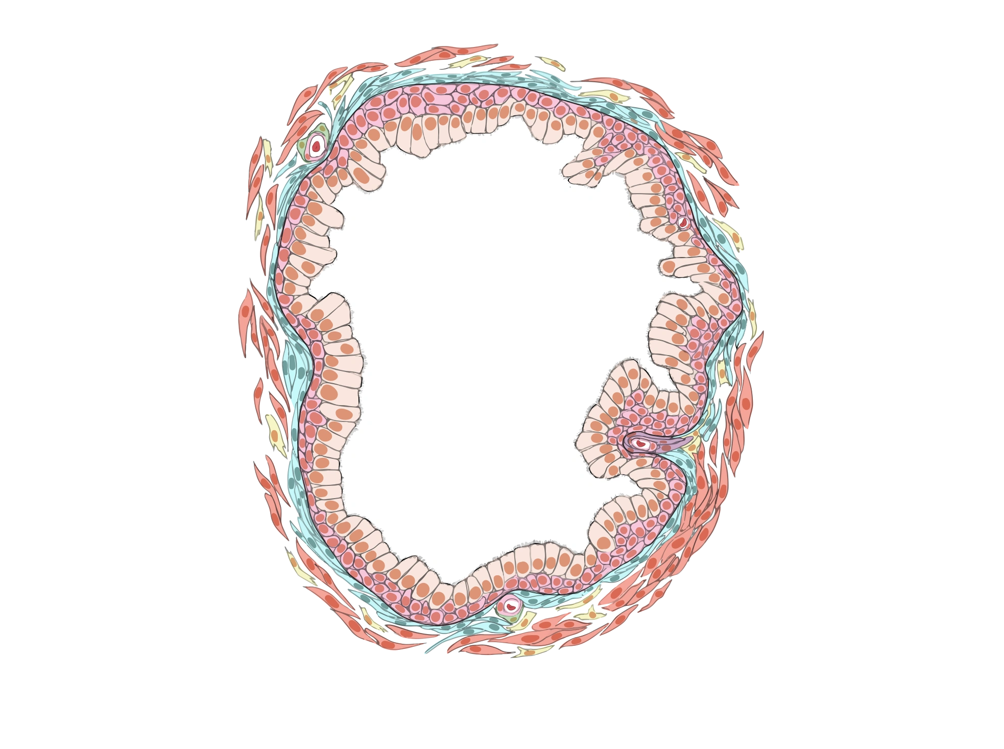 Secretions
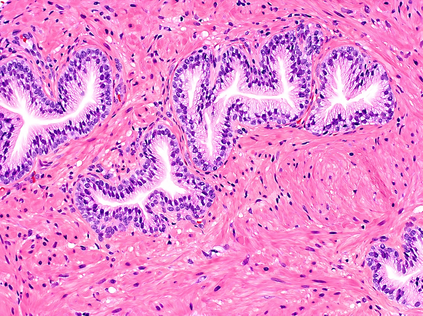
These tiny glands make up the tissue we see under the microscope.
When tissue needs to be checked for cancer, a biopsy takes small samples. They are studied by a pathologist, a doctor who examines tissue under the microscope.
Compare two samples: one of normal tissue and one with cancer.
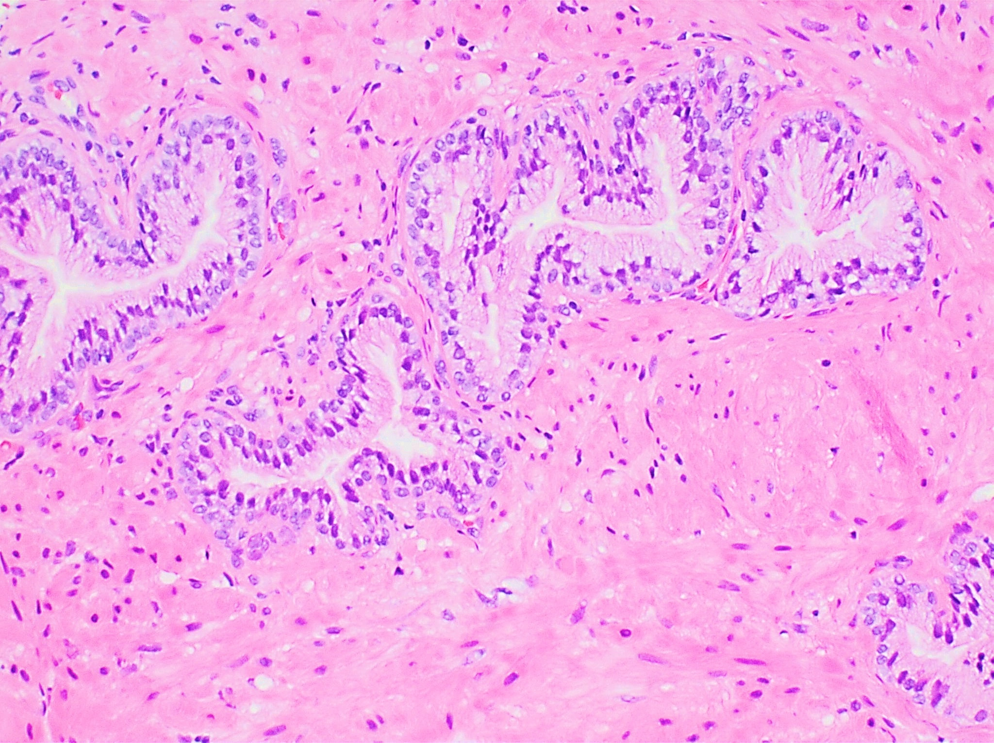
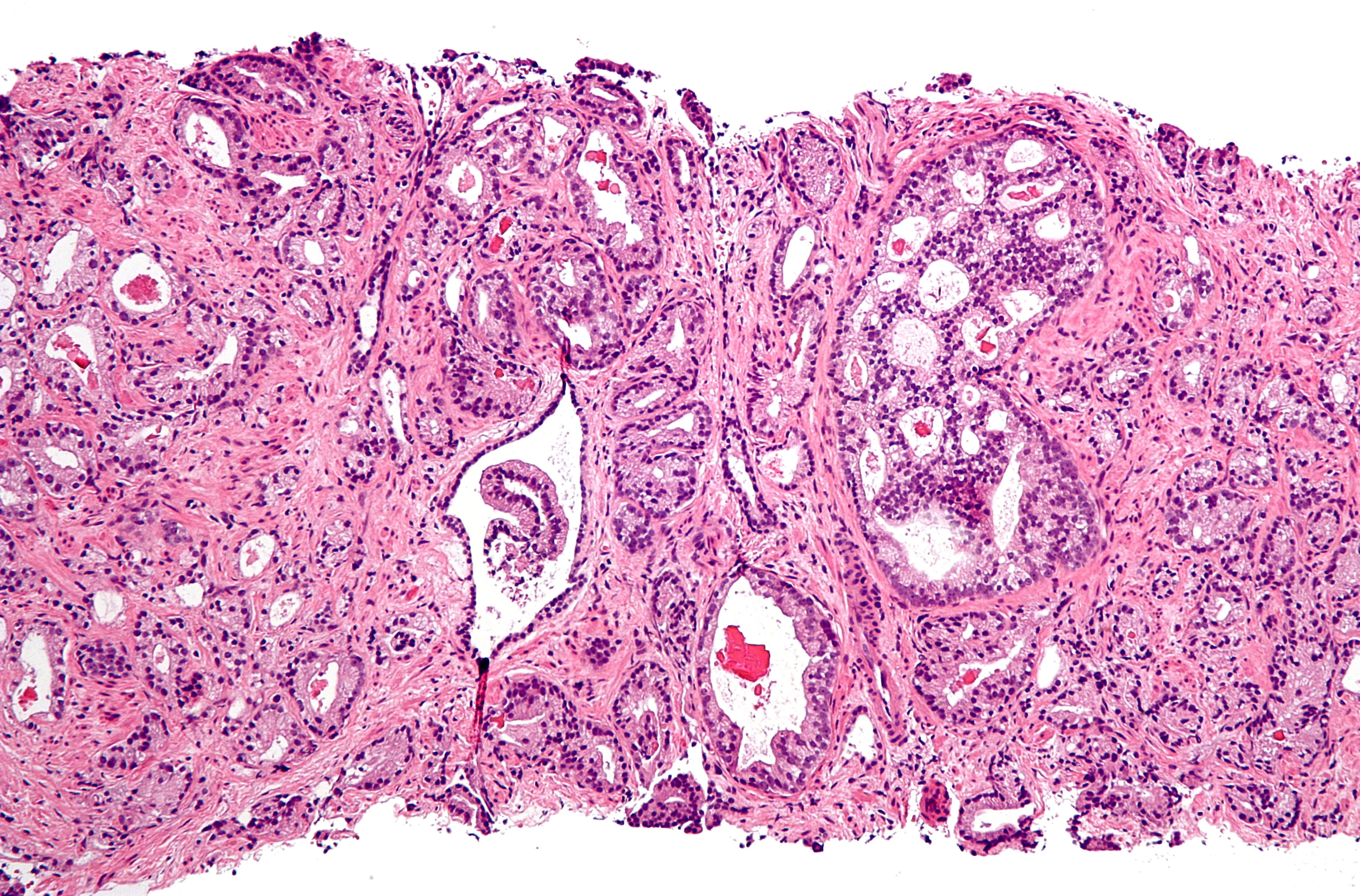
Cells surround pale spaces. Pink tissue keeps the glands apart.
Adenocarcinoma starts in the glands. It can invade, which means it grows into nearby tissue.
In this sample, the glands lose the space between them and merge into irregular groups.
These tissue changes can’t be detected from the strength of the urine stream or the size of the prostate.
Something you can do starting today
Try cutting back on coffee and alcohol, and avoid drinking lots of fluids right before bed. These changes can ease the discomfort.
Write down when the way you urinate started to change. It helps your doctor understand what’s happening and decide what to check.
If you can’t urinate, get medical care right away.
Knowing the cause helps you choose how to ease the symptoms.
I’m your friend, Dr. Carlo De la Sancha. Health and science for everyone.
Official BodyParts3D anatomical render, recolored for the page.
CC BY 4.0National Cancer Institute illustration. The original labels and arrows were removed, the panels were cropped, and labels were added in English.
Public domain; NCI 7138, with restrictions listed as “None” in the record reproduced on Wikimedia CommonsOriginal labels, guide lines and scale bar removed; anatomy, colors and the central space unchanged. Framed for display.
CC BY 4.0Original micrograph, cropped for display; no lettering added and no structures altered. The comparison is about how the glands are organized, not their size.
CC0Original micrograph, cropped for display; no structures altered. The comparison is about how the glands are organized, not their size.
CC BY-SA 3.0CC0 micrograph by Mikael Häggström, MD. The site applied a generative transformation to create the dramatic tissue image; the separate original photographs support the structural comparison.
CC0The microscope images are separate examples of human prostate tissue stained with hematoxylin and eosin. They allow a comparison of how the glands are organized, not of absolute sizes or of changes in one person over time. The original magnifications are not documented in a comparable way.