

H. pylori · Visual journey

H. pylori: how it can change the stomach lining.

From the body to the tissue, to see how inflammation changes the lining.

Begin the journeyScroll to change scale

Organ

The stomach.

Its wall contains the lining: the surface where H. pylori can persist.

Tissue

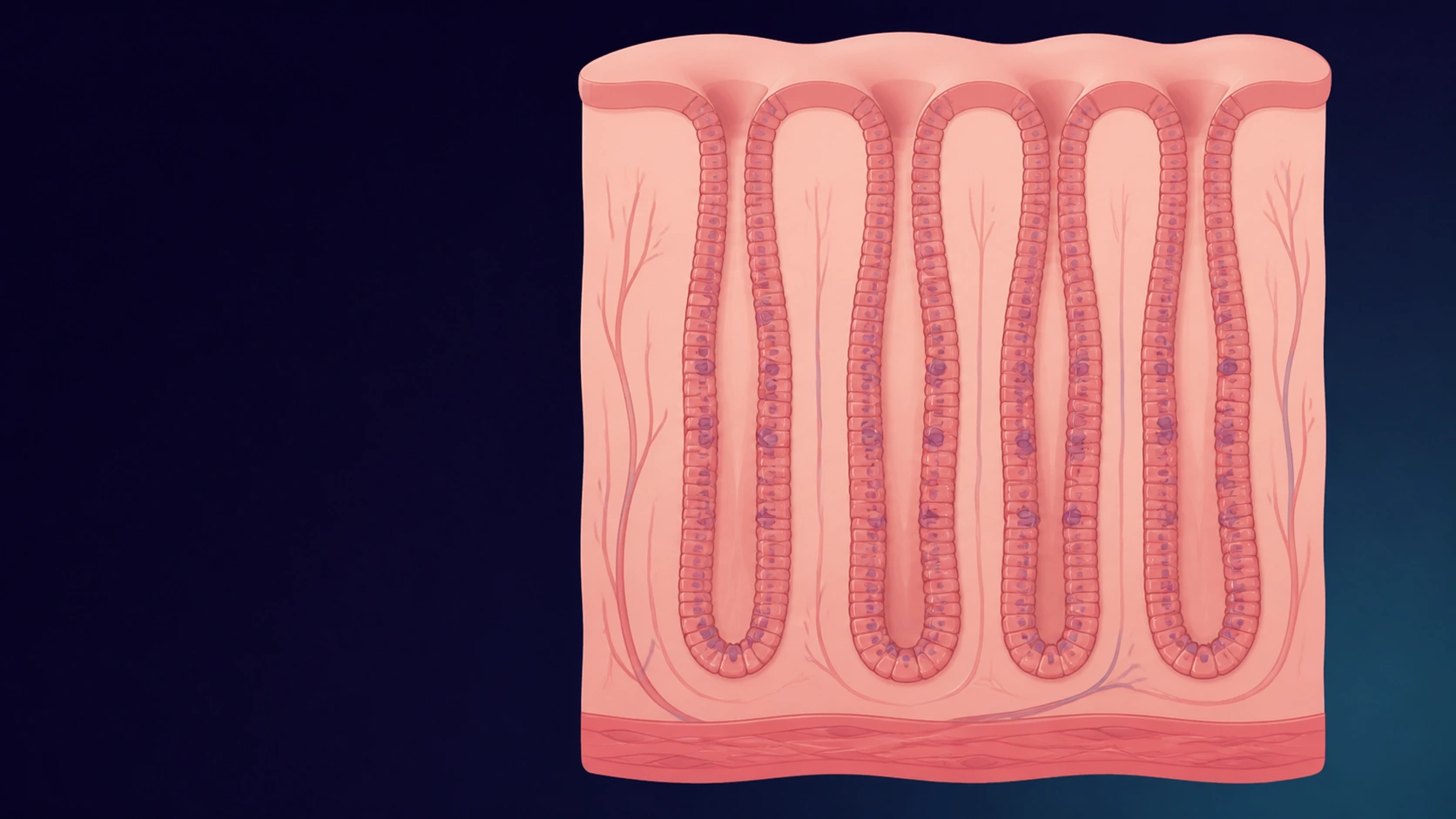
The lining.

Its glands look like tightly packed, orderly tubes. There are very few immune cells between them.

Under the microscope

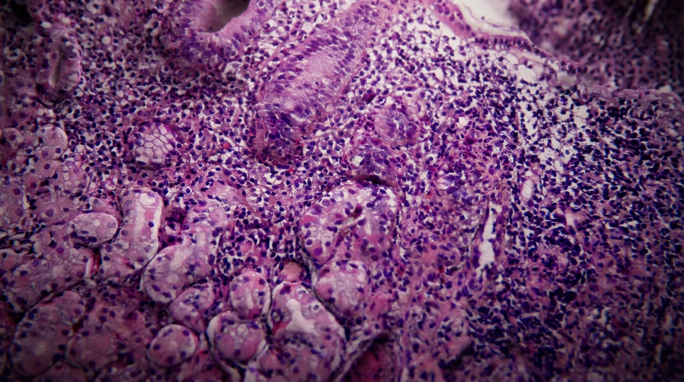
H. pylori gastritis.

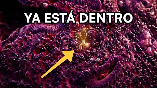
Between the glands, the many small purple dots are immune cells that have accumulated with inflammation.

Compare the tissue

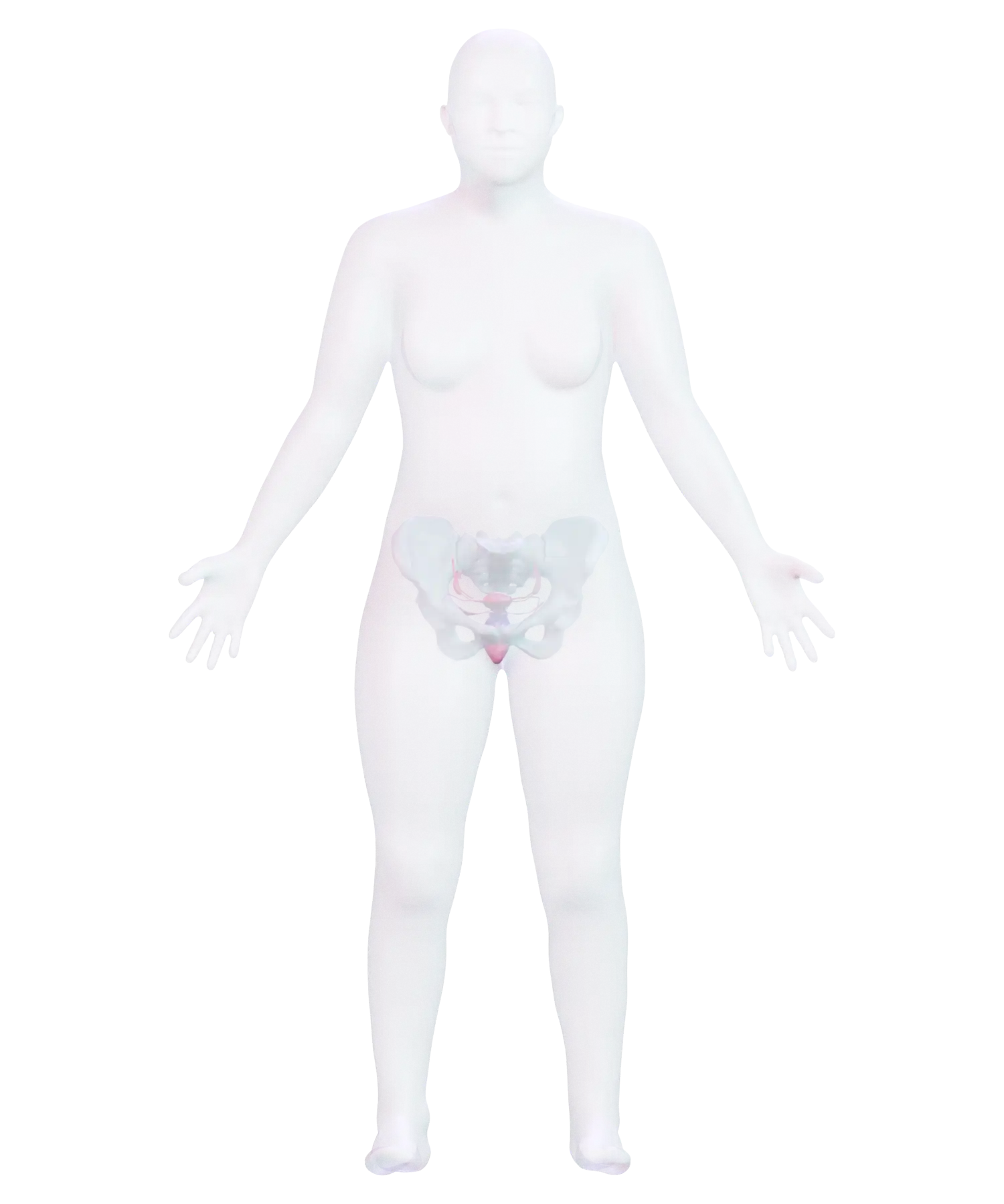
The sequence moves through the body, an anatomical diagram of the stomach, the organization of its lining, and a real biopsy with H. pylori-associated gastritis.

Evidence under the microscope

Look closely at the purple dots.

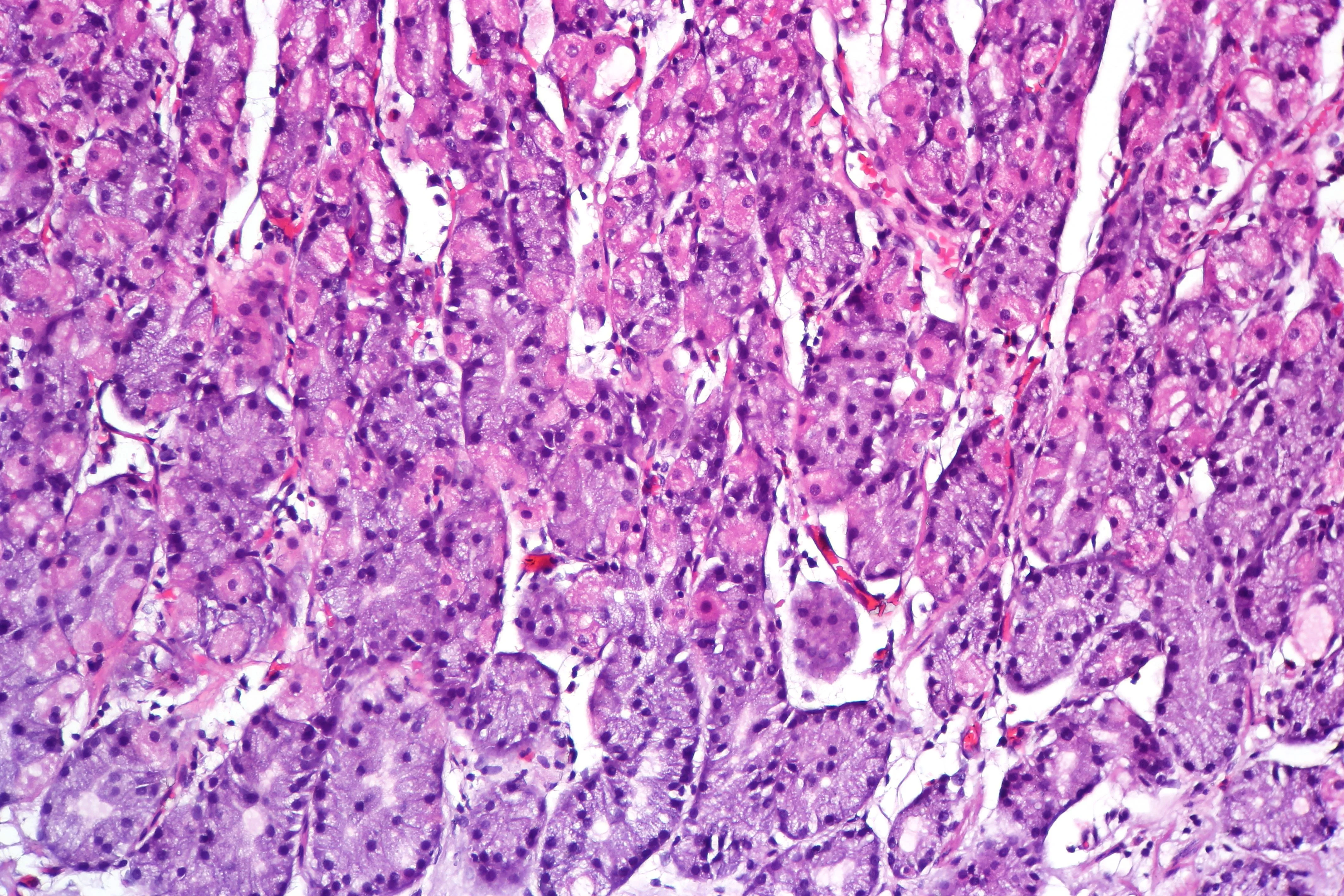
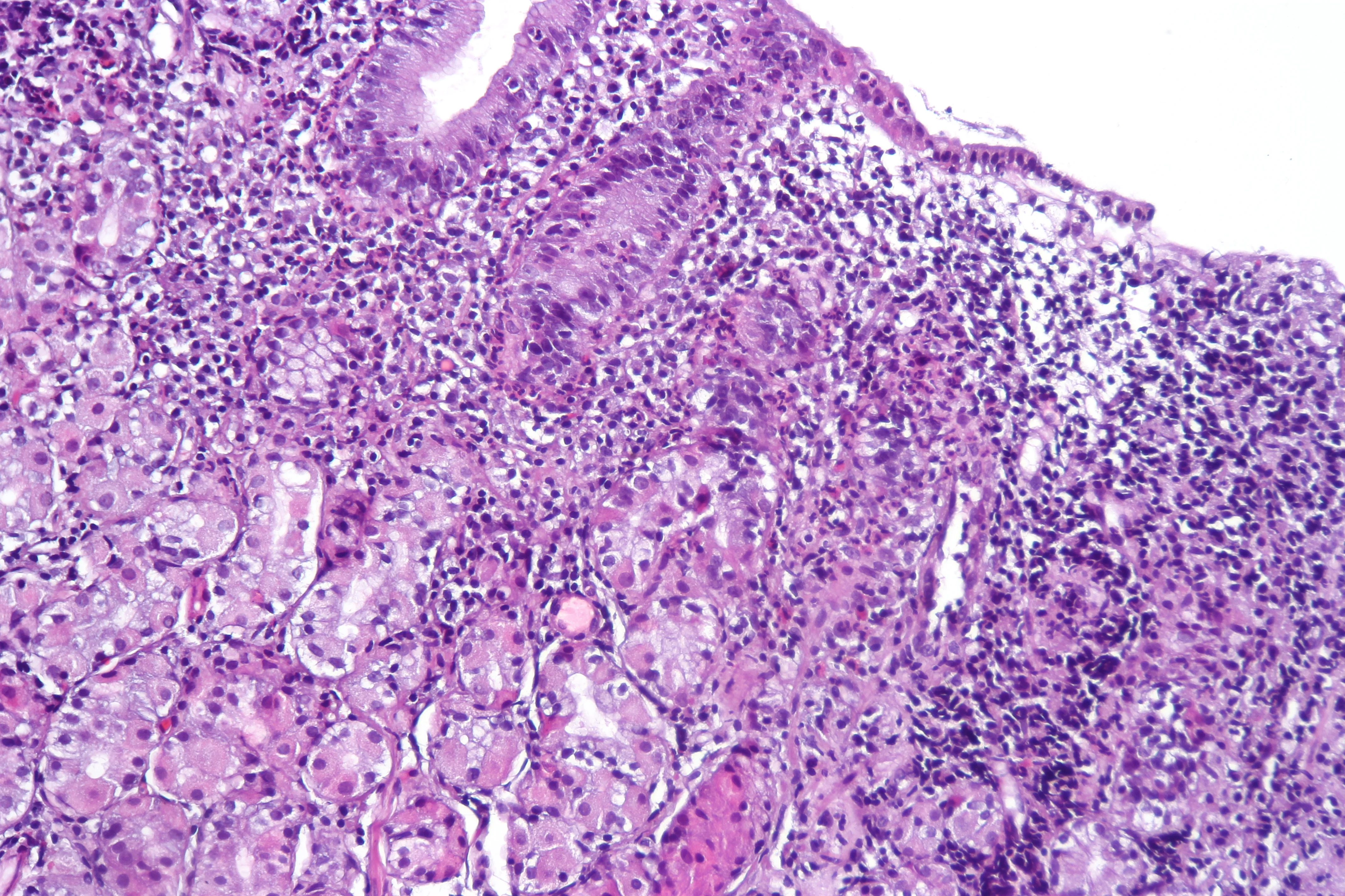
The stain makes the glands pink and the nuclei purple. Look between the glands: when that space fills with small purple dots, lymphocytes, a type of immune cell, are present.

H&E micrograph of gastric oxyntic mucosa without inflammationNo inflammation
Tissue without inflammation

These pink columns are glands. There are few small purple dots between them.

H&E micrograph of gastric mucosa with Helicobacter pylori-associated gastritisWith gastritis
Tissue with gastritis

Now the small purple dots accumulate and fill the space between the glands. They are lymphocytes that arrived with the inflammation.

Over the years, this inflammation can leave a mark on the stomach lining.

What can happen over time

Inflammation can leave a mark.

Chronic H. pylori infection causes gastritis and increases the risk of stomach cancer. Many people with the infection never develop it.

  1. 01Chronic active gastritisImmune cells continue to enter the tissue.
  2. 02AtrophySome glands are lost.
  3. 03Intestinal metaplasiaThe stomach lining begins to resemble the intestine.
  4. 04DysplasiaThe cells lose their organization and grow abnormally.
  5. 05AdenocarcinomaThe cells cross the barrier and invade the tissue.

Free guide

Understand your upper GI biopsy.

A guide to the most common findings in biopsies of the esophagus, stomach, and duodenum.

Request your free guide with your first name and email address.

Get the free guide

You’ll also receive our newsletter every two weeks. You can unsubscribe at any time.

The form opens in Kit. Confirm your email address to download the guide.

Free guide

Your colonoscopy found a polyp

What the polyp means, how to read its report, and when your next colonoscopy is due.

Get the free guide

Know someone who would find this helpful?

Share it with them.