
Bone · Visual journey

Your bones are alive.

They may seem fixed and lifeless. But inside, they hold living cells that renew them. Step into the femur, your thigh bone, to see what’s going on beneath that hard surface.

Scroll to step inside the bone ↓
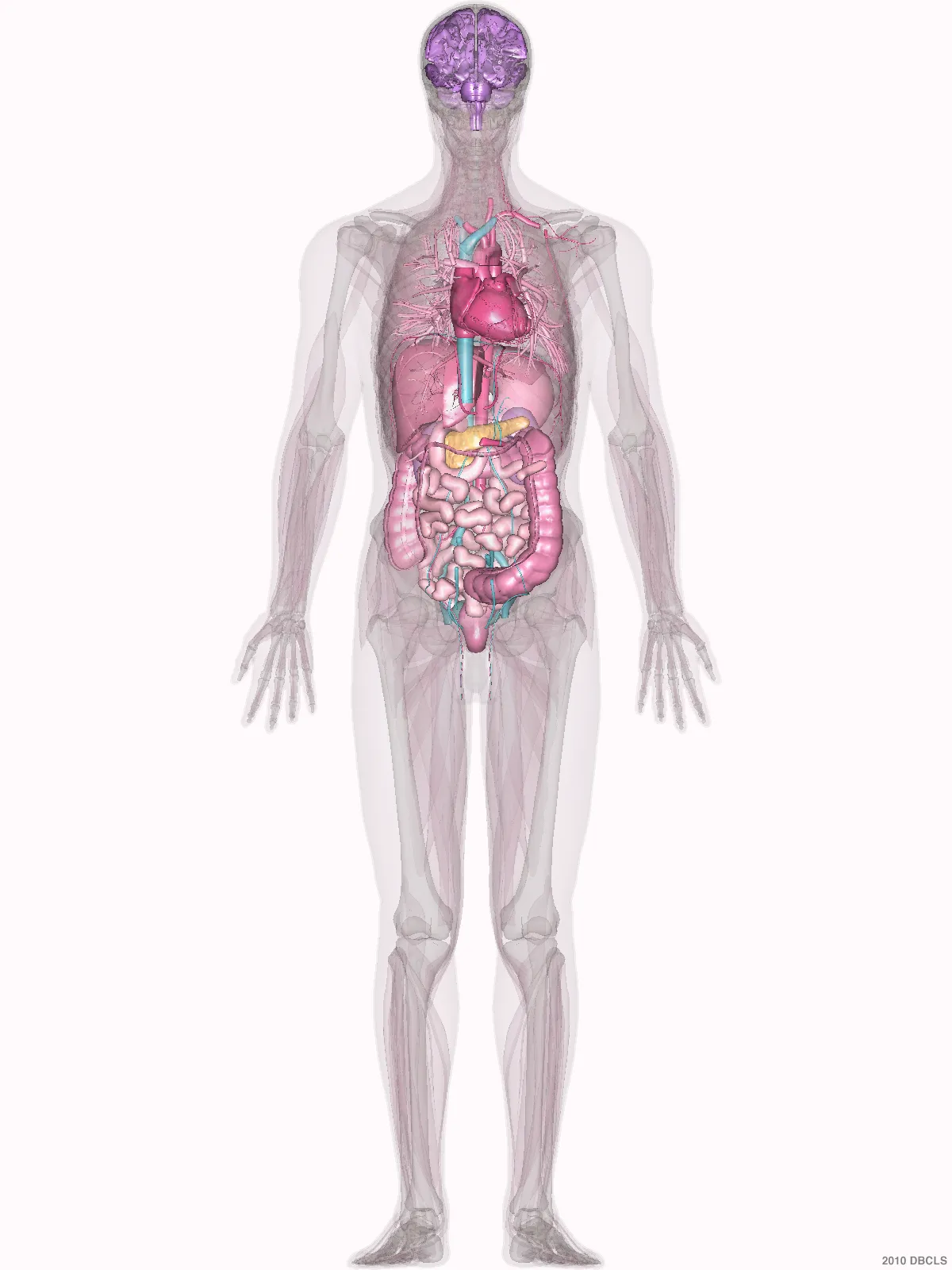
Body figure with the thigh marked

Inside a long bone

It isn’t a solid block.

The outer layer is compact bone, dense and strong. At the ends, spongy bone forms a network of thin plates called trabeculae.
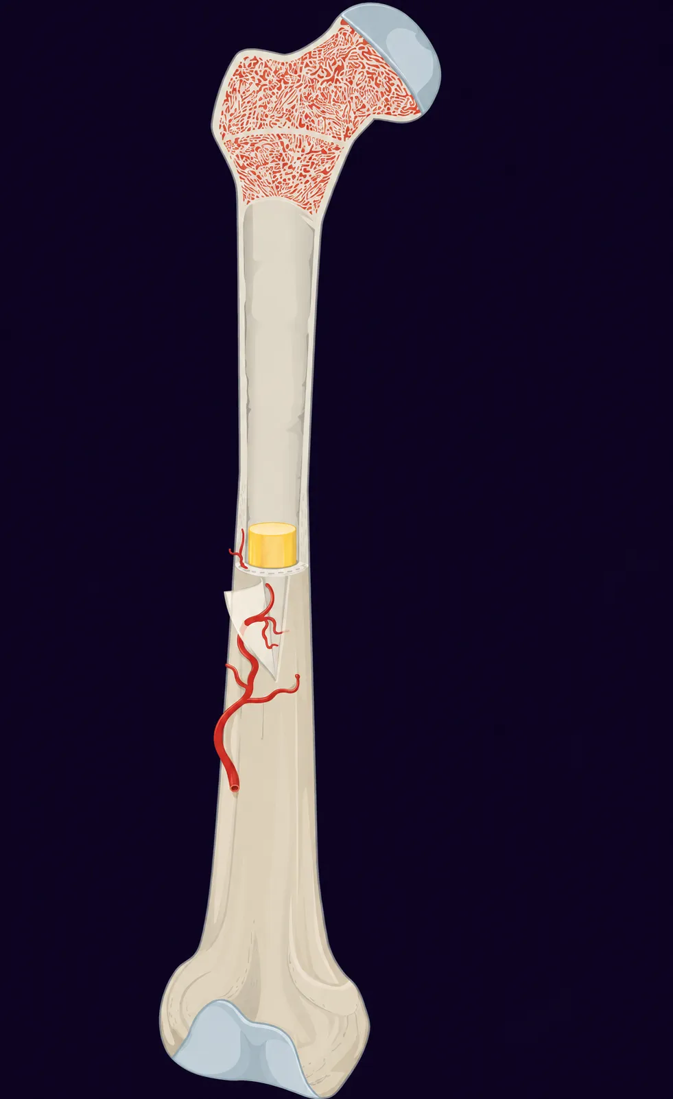
Long bone cut open: a compact outer layer and a spongy network at the ends

The same network, up close

Bone renews itself without losing strength.

In small areas, cells called osteoclasts remove bone. Then osteoblasts lay down a material that hardens as it takes up minerals. This turnover, called remodeling, renews parts of the bone without taking the whole bone apart.

Close-up of the spongy bone in the same illustration

From the tissue to its cells

A cell that builds bone.

The blue cell in the center is an osteoblast. Its nucleus looks purple. It’s one of the cells that form bone, and now you can recognize it under a microscope.
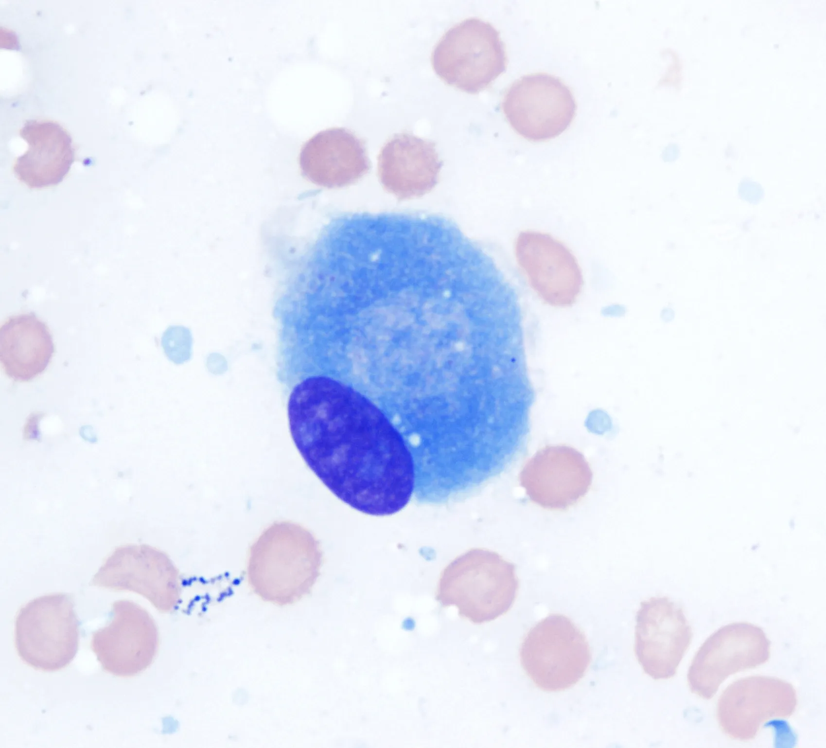
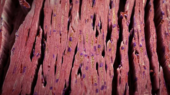
Micrograph of a blue osteoblast in the center with a purple nucleus, without labels or arrows

What you can do

Staying active protects your bones, too.

If, over many cycles, more bone is removed than is formed, bone loses structure. But bone responds to use: physical activity helps keep it strong.

I’m your friend, Dr. Carlo De la Sancha. Health and science for everyone.

This week, set aside time to walk and to do strength exercises suited to your fitness level. Working on your balance also lowers your risk of falls. If you have osteoporosis (a disease that weakens bones) or have had a fracture, ask your doctor how to adapt your exercise before you start.

What else changes in your body over the years?

What aging looks likeExplore how the body changes with age →
Medical sources and images

Images and licenses

Each anatomical illustration keeps its source credit. Original labels were removed where they existed, and backgrounds were adapted to match the page. Close-ups are tighter framings of the same image. Gabriel Caponetti’s micrograph shows an osteoblast with Wright-Giemsa stain at 100×, according to its author. The full original is kept, in color and without a pointer.

Know someone who would find this helpful?

Share it with them.