
Skin · Visual journey

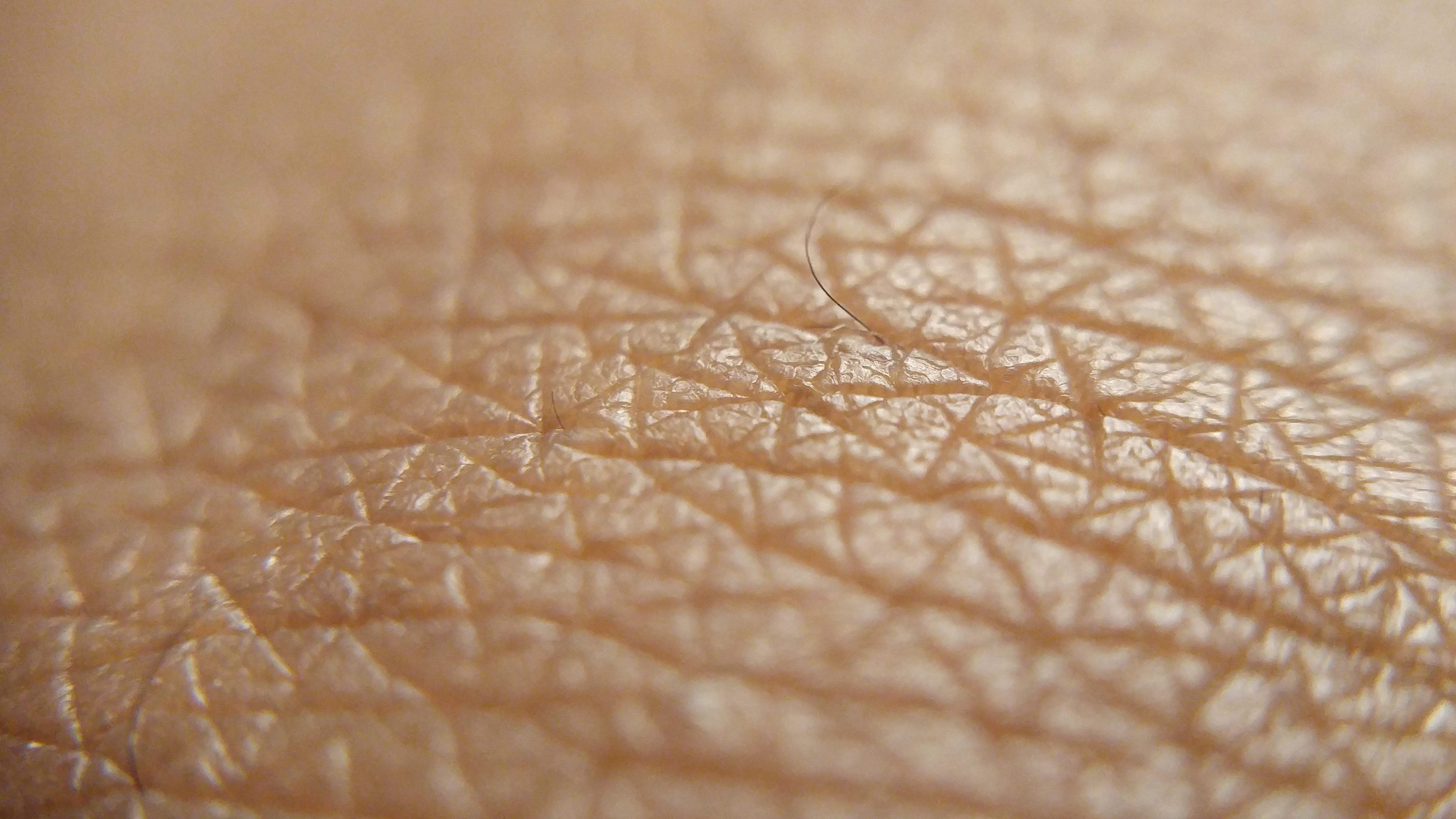
Before the mole, look at the skin.

From the surface you see to the cells that give it color.

Start the journeyScroll to change scale

Tissue

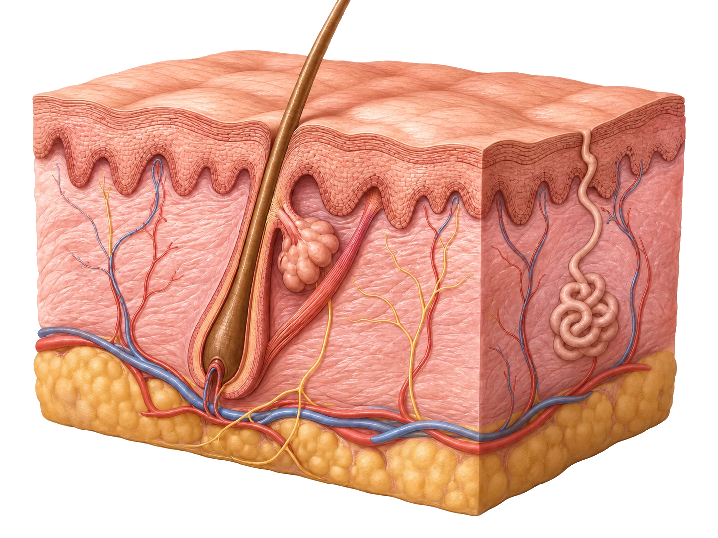
Three layers, many structures.

The epidermis covers. The dermis supports. Beneath them, fat cushions. Vessels, nerves, and follicles run between them.

Cells

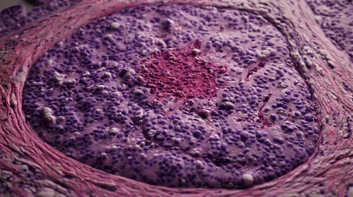
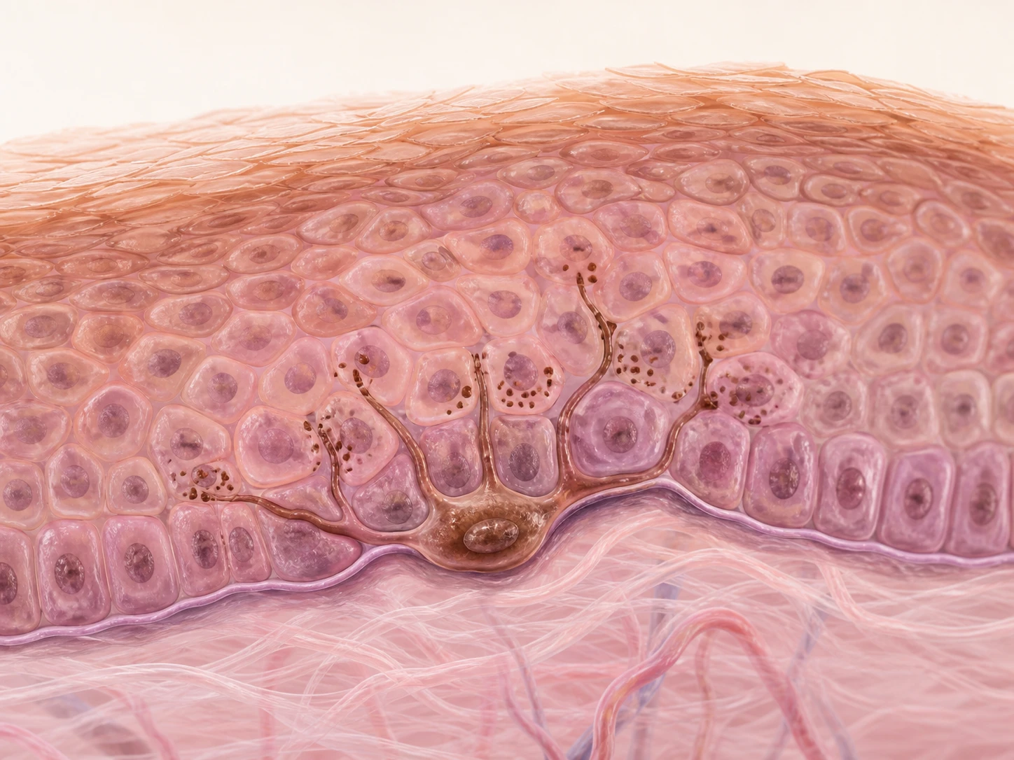
One melanocyte among many cells.

It lives at the base of the epidermis and extends delicate arms to share pigment with neighboring cells.

Under the microscope

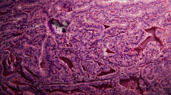
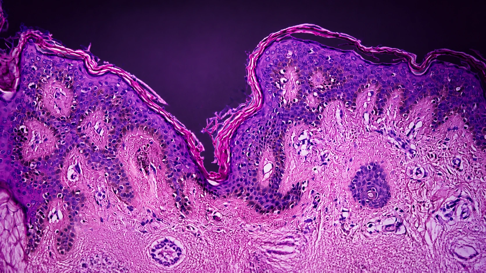
First: normal epidermis.

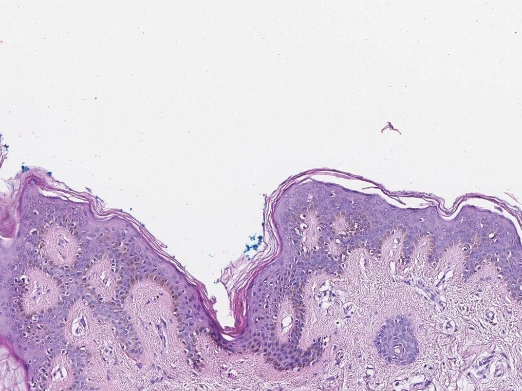
The purple band at the top is the epidermis. Its keratinocytes line up like bricks over the dermis; this will be our reference.

Compare the moles

The sequence begins with a macroscopic photograph of normal skin. It then shows an anatomical section with epidermis, dermis, and subcutaneous tissue, a magnified melanocyte at the junction, and an H&E micrograph of normal epidermis.

At a glance

A common mole and one that deserves evaluation.

No single sign determines whether a lesion is concerning. Asymmetry, irregular borders, several colors, and above all change over time all matter.

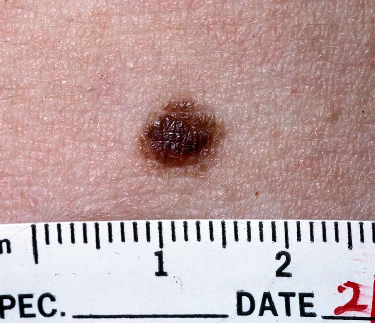
Benign mole that is round, symmetric, and uniform in color
Common mole

Round, symmetric, and fairly uniform in color.

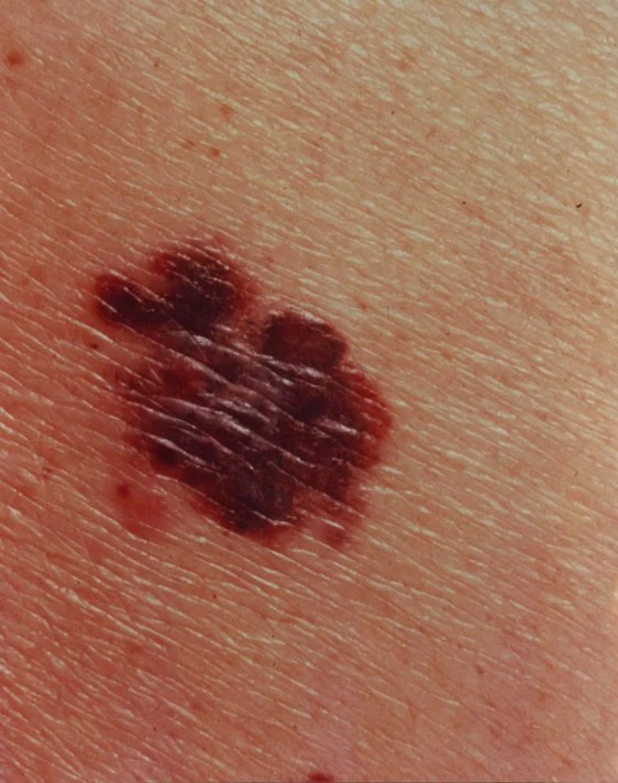
Melanoma with asymmetric shape, irregular borders, and color variation
Suspicious lesion

Asymmetric, with irregular borders and more than one shade. A recent change also matters.

Now, under the microscope

The difference is in the organization.

The same boundary between epidermis and dermis is our guide. First recognize normal skin; then look at where the pigment-producing cells are located.

Normal skin stained with H&E, with orderly epidermis over the dermisThe reference
Unaltered skin

The epidermis forms an orderly band over the dermis. This line will be our point of comparison.

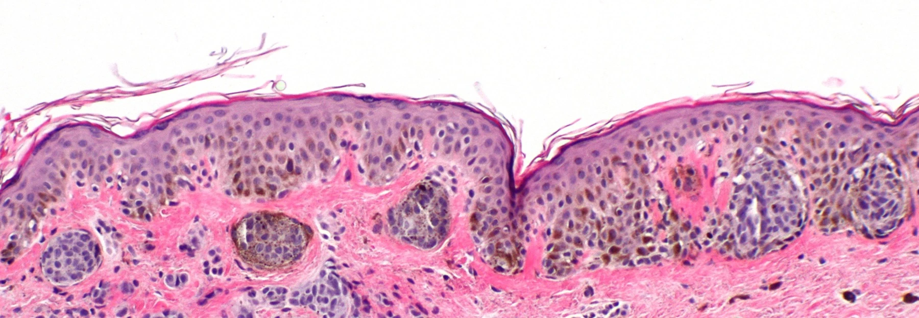
Compound nevus stained with H&E, with melanocyte nests grouped at the junction and in the dermisLower and grouped
Compound nevus

This is a benign mole. The pigment-producing cells form rounded groups along the boundary and below it.

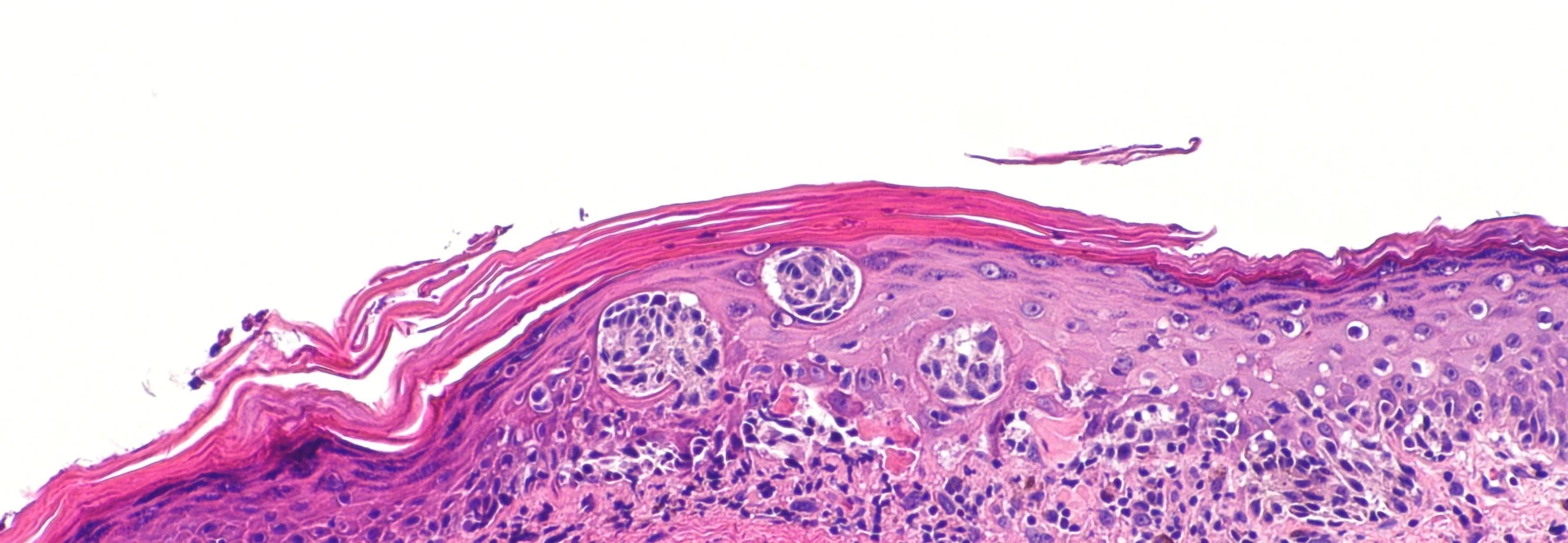
Melanoma in situ stained with H&E, with atypical melanocytes rising through the epidermisScattered and moving upward
Melanoma in situ

This is a cancer still limited to the epidermis. The cells lose their organization and rise among neighboring cells.

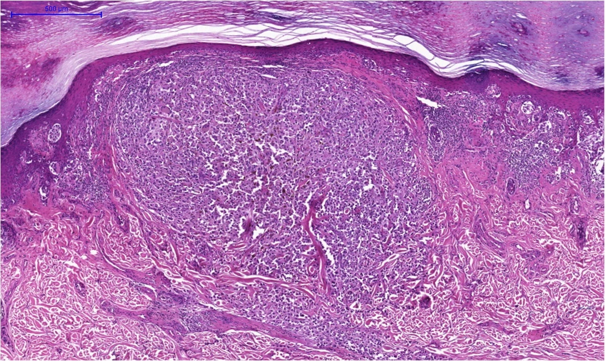
Invasive melanoma stained with H&E, with a nodule of tumor cells extending into the dermisIt has crossed the boundary
Invasive melanoma

Invasive nest: the cancer has crossed the epidermal boundary and forms a nodule within the dermis. That is why it is no longer called “in situ.”

Know someone who would find this helpful?

Share it with them.