
Breast · Visual journey

A lump is not a diagnosis.

From the body to the tissue, down to the boundary that determines the diagnosis.

Begin the journeyScroll to change scale

Organ

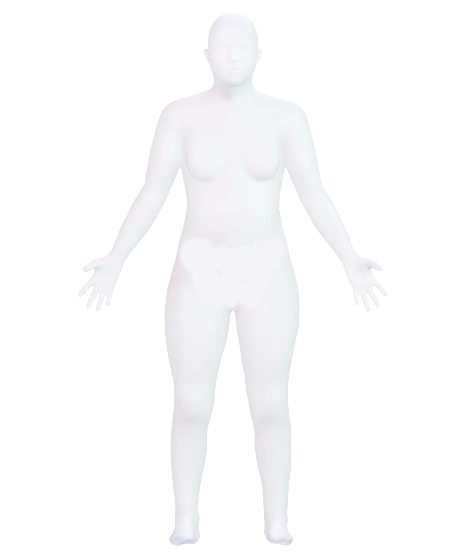
The breast.

Inside, it resembles a tree: ducts lead to the nipple, with milk-producing lobules at their ends.

Tissue

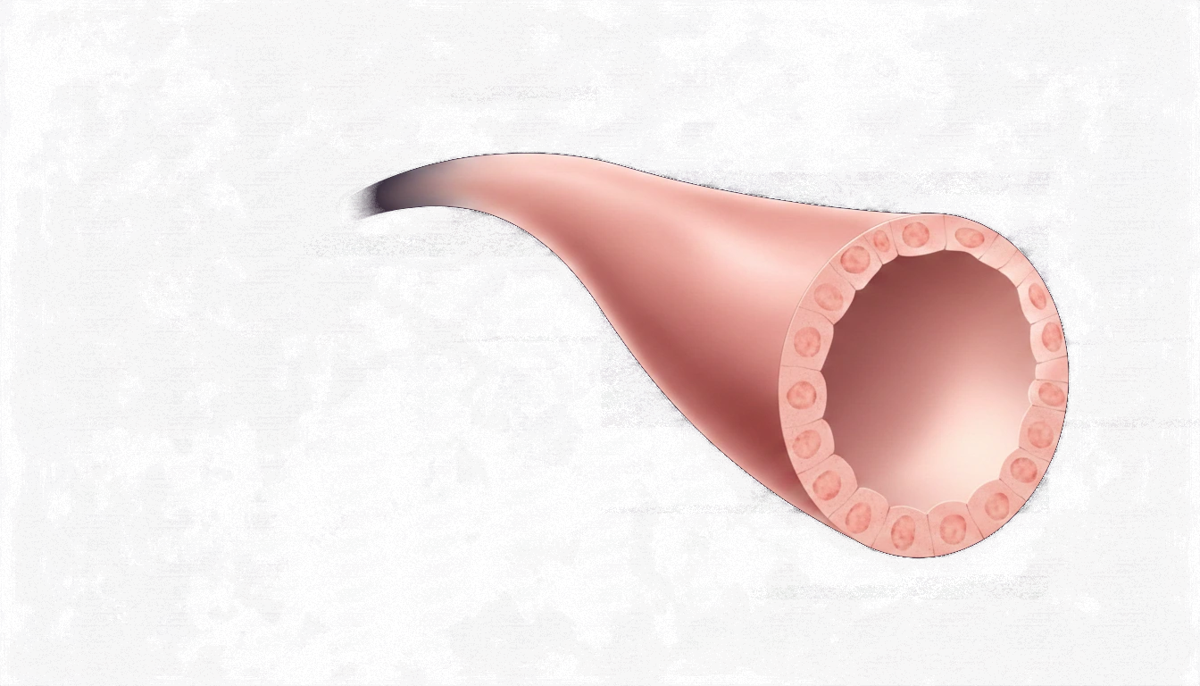
The duct.

Each duct is a tube with its own wall. That wall separates what happens inside from the rest of the breast tissue.

Under the microscope

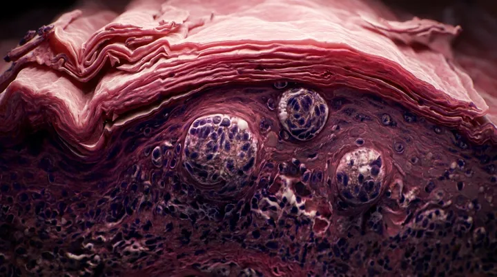
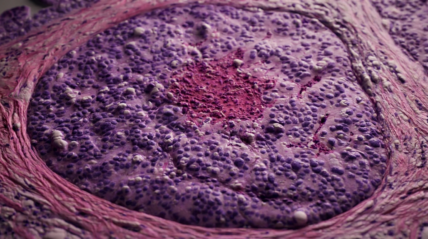
Ductal carcinoma in situ.

Abnormal cells fill the duct and reach its boundary. On the other side of the wall, the tissue remains intact.

Compare the tissue

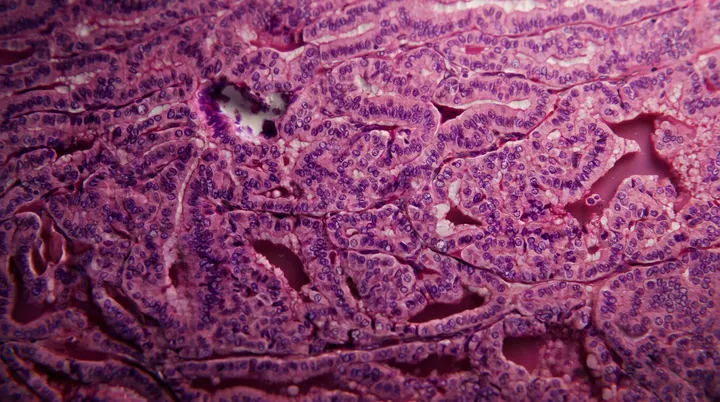
The sequence moves through the body, an anatomical diagram of the breast with its ducts and lobules, a duct viewed from the inside with its wall, and a real biopsy with ductal carcinoma in situ.

Evidence under the microscope

Look closely at the edge of the duct.

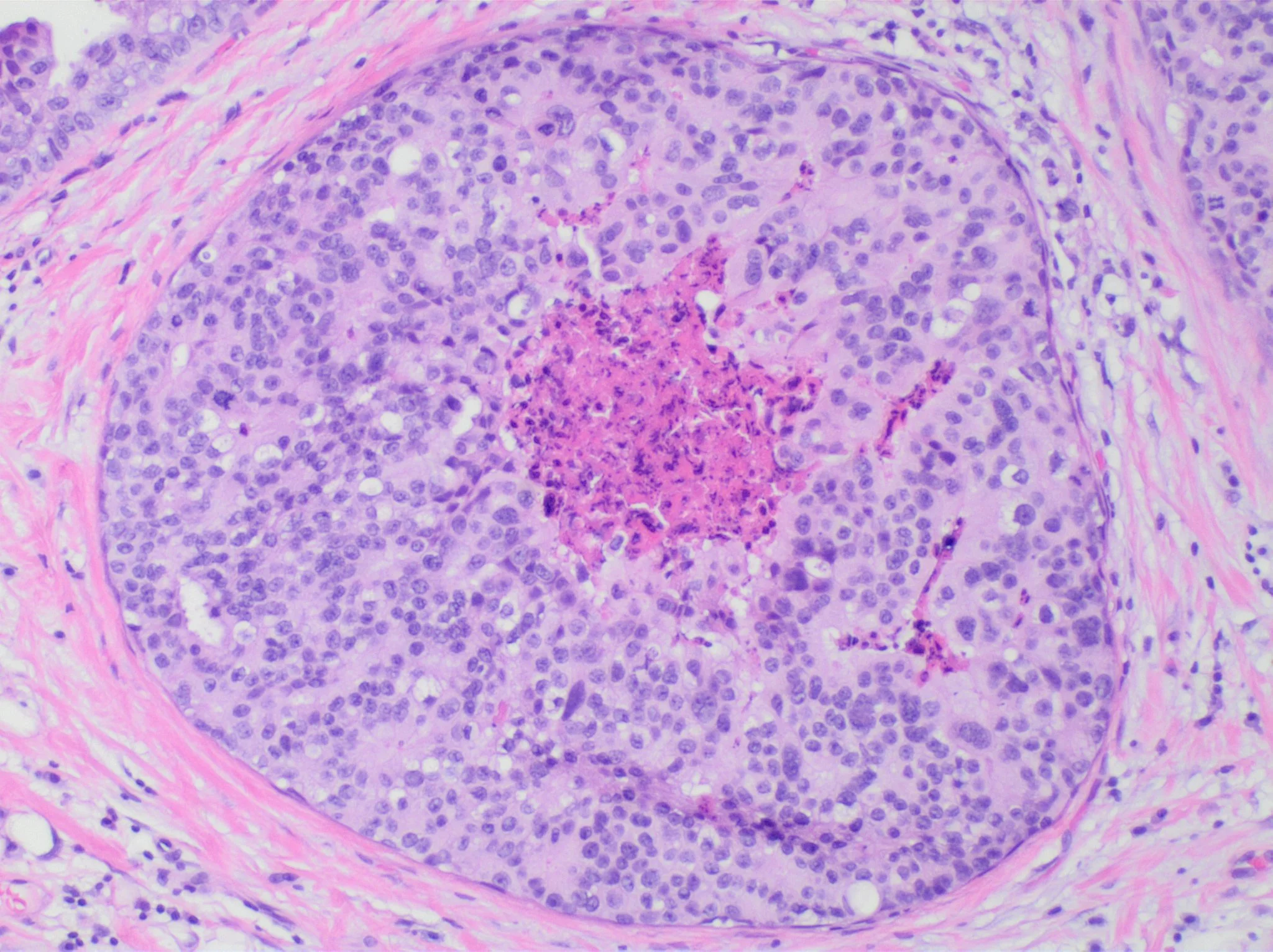
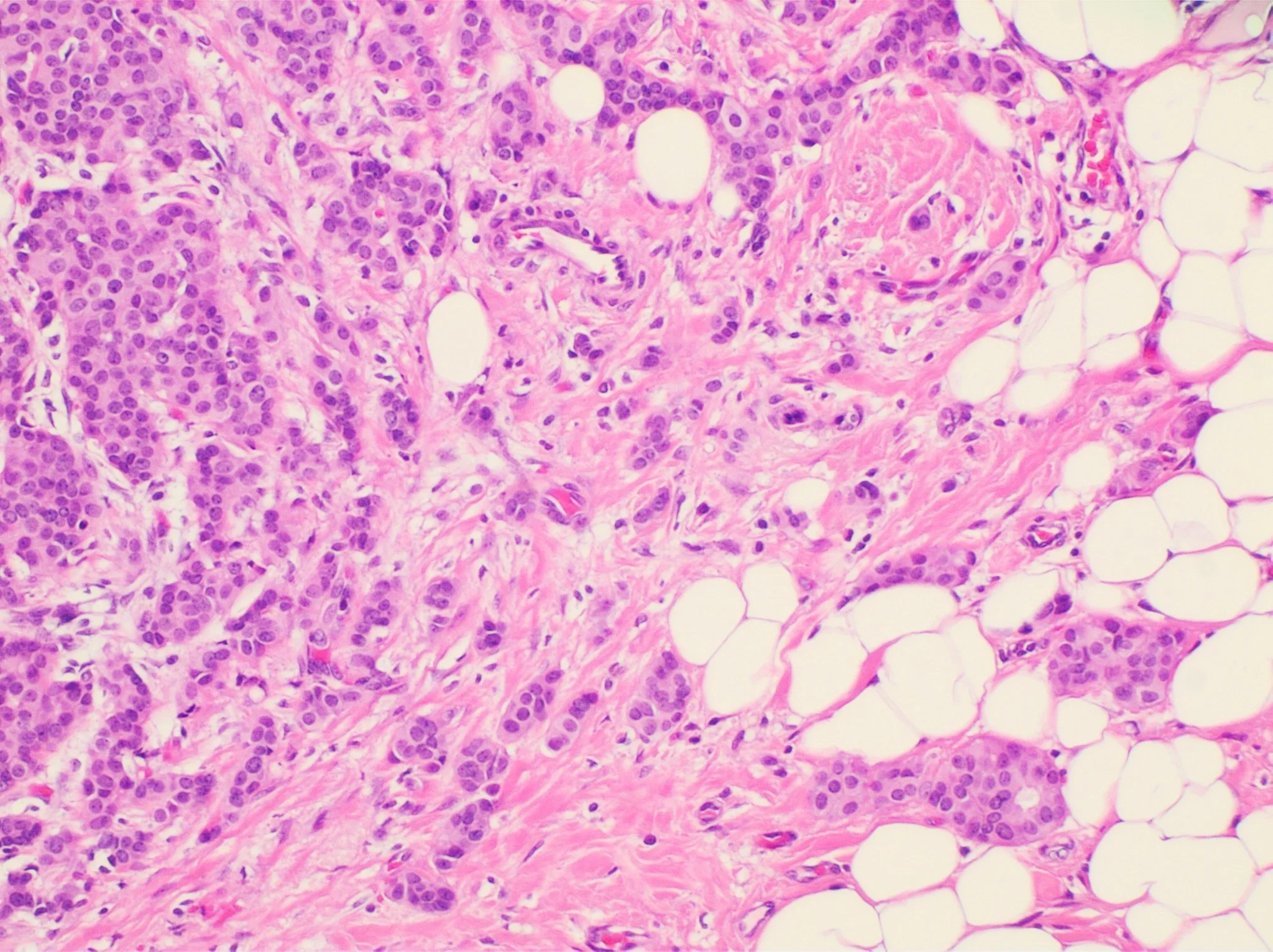
The stain makes the supporting breast tissue pink and the cell nuclei purple. Look for one thing: whether the purple cells stay inside the circle or appear outside it.

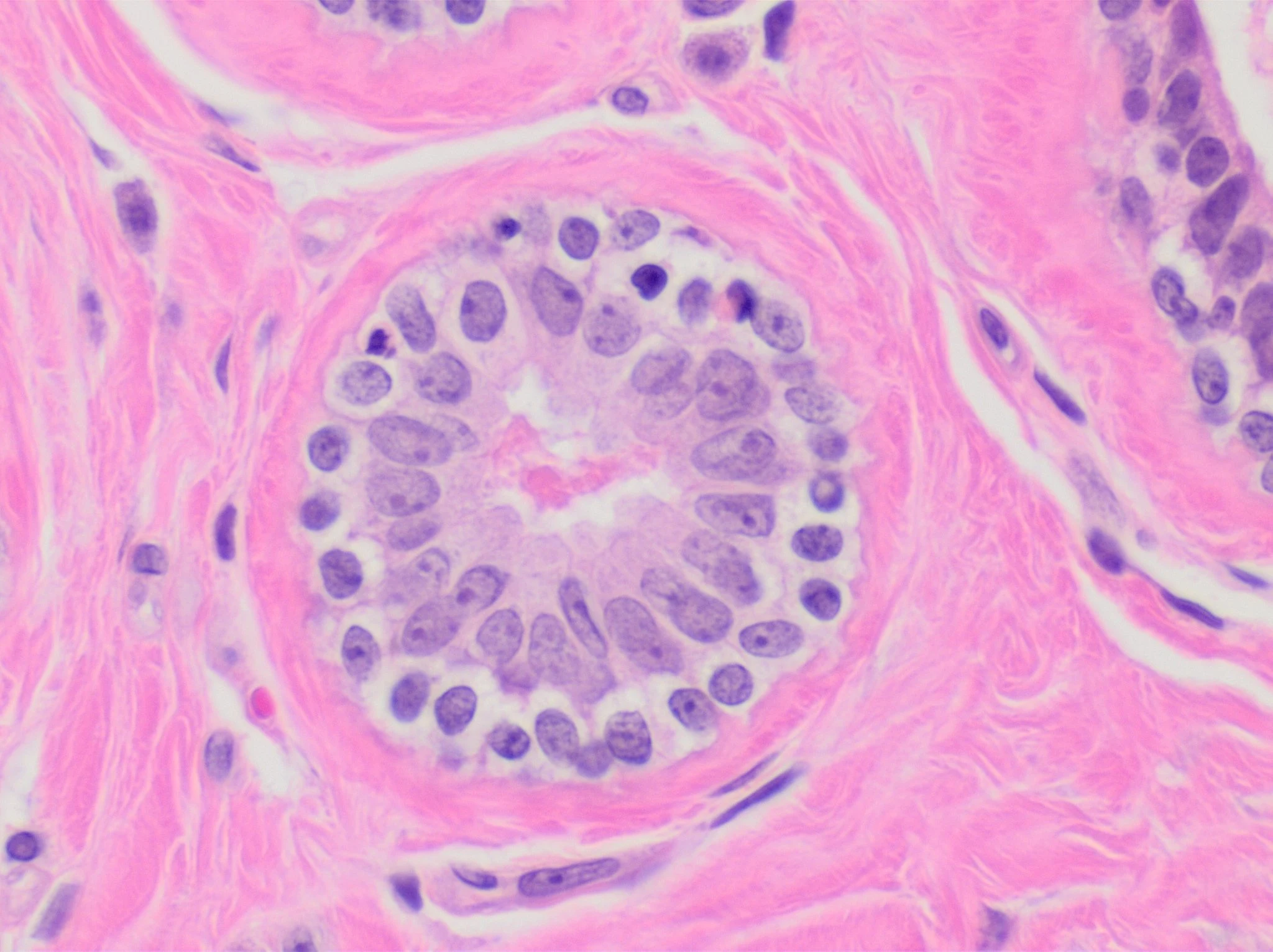
H&E micrograph of an unremarkable breast acinus, with orderly cells surrounding an open lumenUnremarkable
First: normal breast tissue

The cells form an orderly layer around an open center. This is the reference for recognizing what changes next.

H&E micrograph of a breast duct filled by ductal carcinoma in situInside the duct
Ductal carcinoma in situ

The purple cells fill the circle up to its boundary and stop there. The surrounding pink tissue remains clear of them.

H&E micrograph of invasive ductal carcinoma with groups of cells scattered through breast tissueOutside the duct
Invasive carcinoma

There is no longer a circle containing them: groups of purple cells are scattered through the pink tissue and fat.

That boundary determines the words that will appear in the report.

Free guide

How to understand your breast biopsy report.

Help understanding the terms in your report, with questions to discuss with your doctor.

Request your free guide with your first name and email address.

Get the free guide

You’ll also receive our newsletter every two weeks. You can unsubscribe at any time.

The form opens in Kit. Confirm your email address to download the guide.
Medical transparencySources and scope

Medical evidence, image provenance, and the editorial transformations applied.

The anatomical illustrations were cleaned to show one idea per frame: labels and guides were removed, and the anatomy was not redrawn. The dramatic reveal is based on the cited biopsy; the immediate comparison uses the original micrographs with tighter framing.

This journey explains how a report is read; it does not replace review of your own report. The terms are broadly consistent, but their meaning depends on the complete case. Take your report to your appointment.

Sources verified through August 22, 2026.

Know someone who would find this helpful?

Share it with them.