
Intestines · Visual journey

Crohn’s disease and ulcerative colitis: when the bowel is inflamed from within.

Both are inflammatory bowel diseases, in which the immune system inflames the digestive tract, with flare-ups and remissions that can last for weeks or years.

But they are two different diseases, not two stages of the same one. Three questions tell them apart: where they strike, how deep they go, and what the pathologist sees in the biopsy.
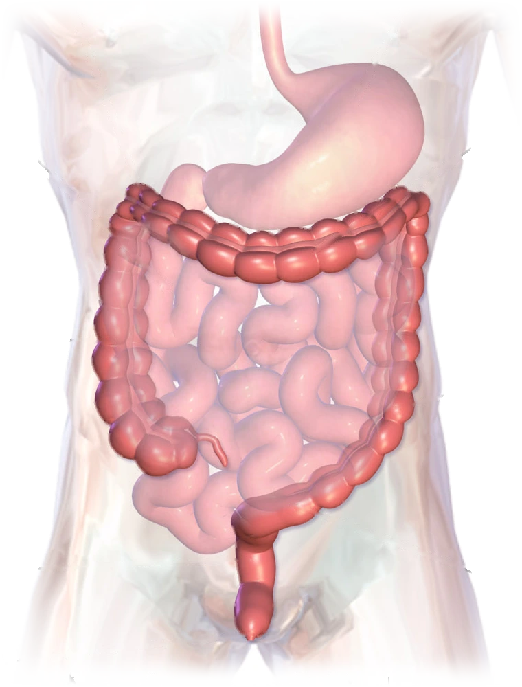
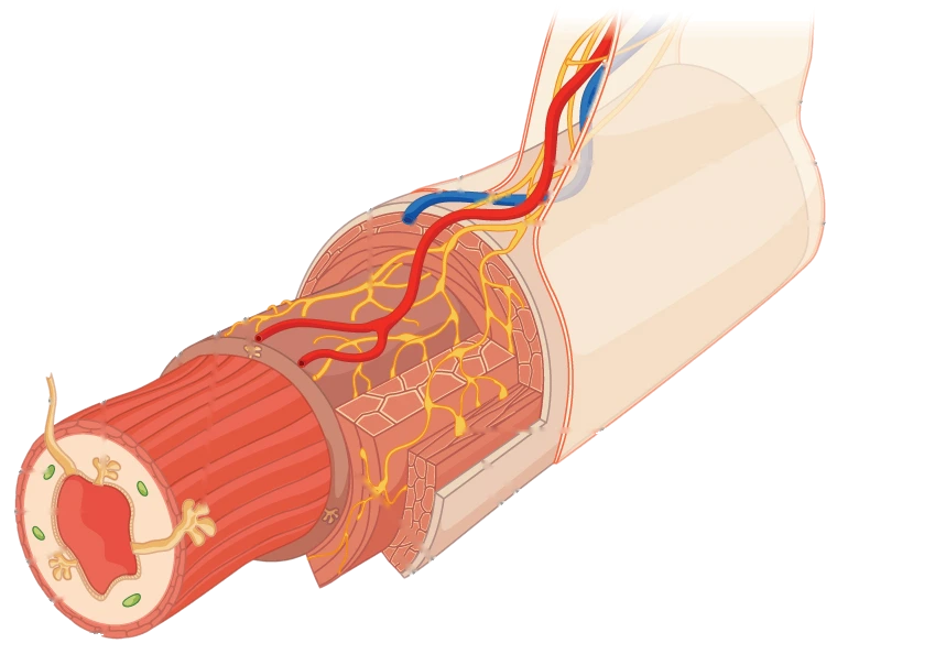
Illustration of the abdomen: the large intestine, in red, frames the loops of the small intestine and runs down the middle to the rectum and anusColonAbsorbs water and forms stoolRectumThe final stretch

Large intestine

The colon and rectum: the final stretch.

The colon frames the abdomen. It absorbs almost all the water and forms stool. Just before the anus comes the rectum.

Diagram of a cut section of the digestive tract: the pale mucosa on the inside, around the central opening, then the red muscle layer, and the serosa on the outsideMucosaThe innermost layerSerosaThe outermost layer

The wall

A wall made of four layers.

Innermost is the mucosa, which touches everything that passes through. Beneath it lie the submucosa and the muscle layer, and on the outside, the serosa.

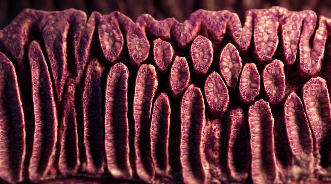
Healthy colon mucosa under the microscope: long crypts lined up in parallel rows, like test tubes

Under the microscope

Healthy mucosa, up close.

The colon’s mucosa is arranged in crypts: small, straight tubes standing side by side, like test tubes in a rack.

Compare where each one strikes

Question 1 · Where

Where each one strikes.

The same map, once for each disease. How far the disease reaches varies from person to person.

Ulcerative colitis

It starts in the rectum and moves up in one continuous stretch.

Illustration of the abdomen showing the large intestine in red, the loops of the small intestine and the rectumRectumIt starts here

It can stay in the rectum, or it can spread up the colon with no healthy stretches in between, until it covers the whole colon.

It doesn’t go beyond the large intestine.

Crohn’s disease

It shows up in patches, anywhere from mouth to anus.

Illustration of the abdomen showing the large intestine in red, the loops of the small intestine and the rectumWhere the small intestine meets the colonThe most common site

It most often affects the end of the small intestine and the start of the colon. But it can appear anywhere from the mouth to the anus.

It comes in patches: inflamed areas with healthy stretches in between.

Question 2 · How deep

How deep it goes into the wall.

The same wall, from mucosa to serosa, once for each disease.

Ulcerative colitis

It stays in the mucosa.

Diagram of a cut section of the digestive tract, showing its layers from the inside outMucosaAlmost always stops here

It inflames the mucosa, the inner lining, and causes ulcers in it.

It usually doesn’t go past the inner layers.

Crohn’s disease

It can go through the entire wall.

Diagram of a cut section of the digestive tract, showing its layers from the inside outFrom the mucosaAll the way to the serosa

It tends to go deeper: it can cross the full thickness of the wall. This is called transmural inflammation.

Over the years, it can narrow the bowel (a stricture) or open a tunnel to the skin or the bladder (a fistula). Treating it well lowers that risk.

What reaches the microscope

A biopsy is a tiny piece of that wall.

A pathologist reads it under the microscope. It’s taken during a colonoscopy: a tube with a camera travels through the rectum and colon and removes tiny pieces of the lining. You don’t feel it.

Because the pieces come from the lining, the biopsy mostly shows the mucosa: what Crohn’s does deeper in the wall may not show up.

Question 3 · What the pathologist sees

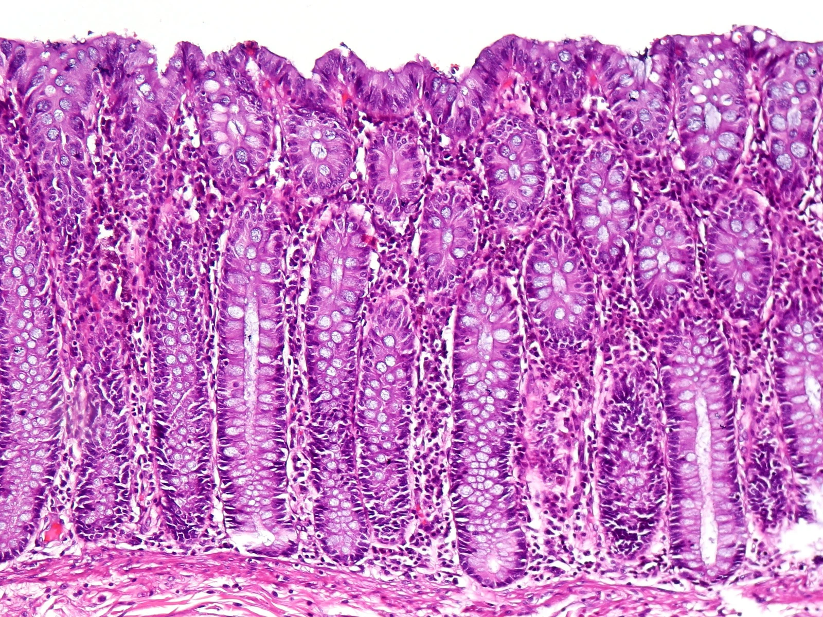
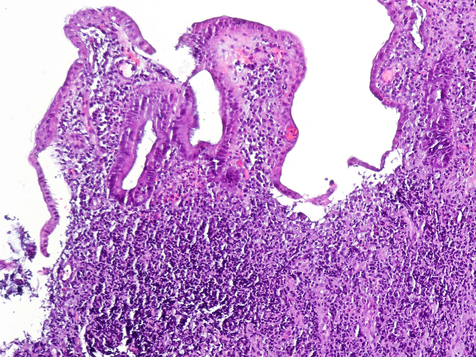
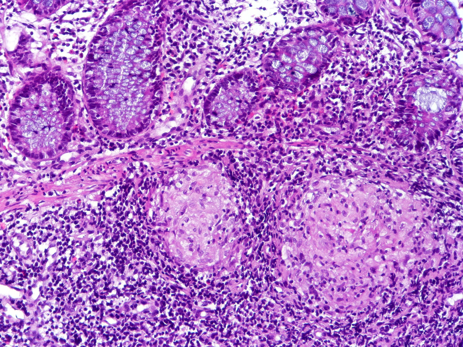
Three colon biopsies under the same microscope.

Biopsies are taken from each section of the bowel, even if it looks healthy, and the samples from each site go into their own jar.

They’re stained with hematoxylin and eosin (H&E): cell nuclei turn purple, and everything else turns pink.

Healthy colon mucosa under the microscope, stained purple and pink: long, straight, parallel crypts lined up side by side

Crypt: a straight tube, in line with the others

Parallel crypts running from the surface down to the base, all the same size and evenly spaced.

Colon mucosa with ulcerative colitis under the microscope: an irregular surface, a long, misshapen crypt and, below it, a mass of inflammatory cells

Distorted crypt

Severe active colitis: an ulcerated surface, crypts that have lost their shape and order, and mucosa packed with immune cells.

Colon biopsy with Crohn’s disease under the microscope: crypts cut crosswise at the top and, below them, two round, pale clusters of cells ringed by dark cells

Granuloma: a cluster of macrophages

Crypts at the top; below, two granulomas: pale clusters of macrophages surrounded by lymphocytes, two kinds of immune cells.

With long-term inflammation, the crypts branch and lose their shape, more so in ulcerative colitis than in Crohn’s.

A granuloma points to Crohn’s, but most biopsies from people with Crohn’s don’t have one, and intestinal tuberculosis can cause granulomas too.

No single image is enough. The diagnosis brings together symptoms, the colonoscopy, where each biopsy came from, lab tests and, when needed, imaging.

What you can control

Know the signs, and come to your appointment prepared.

Neither Crohn’s disease nor ulcerative colitis can be cured with medication, but treatment can give you long stretches without symptoms. And because no single test can diagnose them, what you tell your doctor is part of the diagnosis.

What you can do

Before your appointment, write down your symptoms.

How often you have bowel movements, whether you have to get up at night to go, any blood or mucus, where it hurts, any weight loss, and how long this has been going on. Also note your medications, whether you smoke, and whether anyone in your family has Crohn’s or colitis.

If you’ve already been diagnosed, also keep track of what you eat, changes to your medications and your stress, so you can see what makes your symptoms worse.

After a colonoscopy, ask which parts of the bowel the biopsies came from and what the pathologist saw.

How it’s diagnosed

Several tests, not just one.

Blood tests (for anemia and inflammation), stool tests (for infections and calprotectin), a colonoscopy with biopsies and, sometimes, a CT scan or MRI.

What a colonoscopy is like

Bowel prep, sedation and a ride home.

A special diet and a laxative in the days before, sedation through an IV, and less than an hour for the procedure. Bring someone with you, because you won’t be able to drive. Biopsy results take a few days.

Treatment

It’s tailored to you.

Your gastroenterologist chooses it based on your symptoms and which part of your bowel is inflamed. Keep taking your medications even when you feel well, and check with your doctor before using ibuprofen or any other anti-inflammatory drug.

If you have Crohn’s and smoke

Quitting helps.

Smoking raises the risk of flare-ups and can double the risk of developing Crohn’s. Quitting helps bring the disease under control.

Keeping watch on the colon

Ask when you’re due for a surveillance colonoscopy.

Years of inflammation in the colon raise the risk of colorectal cancer. Surveillance usually starts about eight years after the disease begins and is repeated based on your risk.

You’re not alone

Connect with other people living with the same condition.

Support groups, in person or online, can help you get through flare-ups.

Your friend, Dr. Carlo De la Sancha. Health and science for everyone.

Keep learning

What did the pathologist find in the biopsy of a patient whose ulcerative colitis got worse?

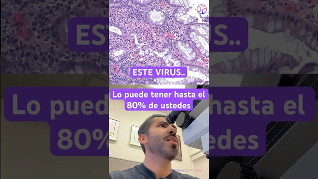
The virus that lies dormant in 80% of you right nowWatch the video about the virus that woke up in a patient with colitis
Medical transparencySources, images and licenses

Where the information comes from

Images

In the diagrams, the original labels and their leader lines were erased and the white background was made transparent; none of the anatomy was redrawn. The arrows and labels you see were added by this site. The opening microscope image is a version of the cited micrograph with more depth. The three biopsy images are crops, with their brightness evened out so they can be compared.

Educational content for the general public. It doesn’t diagnose Crohn’s disease or ulcerative colitis, and it doesn’t recommend treatments: the diagnosis and any decision about medications are up to your health care team.

Know someone who would find this helpful?

Share it with them.